PUBLICATIONS
PUBLICATIONS
Tissue Engineering Part A: Research Advances | September 2023
A Ready-to-Use Purified Exosome Product for Volumetric Muscle Loss and Functional Recovery
Large skeletal muscle defects owing to trauma or following tumor extirpation can result in substantial functional impairment. Purified exosomes are now available clinically and have been used for wound healing. The objective of this study was to evaluate the regenerative capacity of commercially available exosomes on an animal model of volumetric muscle loss (VML) and its potential translation to human muscle injury. An established VML rat model was used. In the in vitro experiment, rat myoblasts were isolated and cocultured with 5% purified exosome product (PEP) to validate uptake. Myoblast proliferation and migration was evaluated with increasing concentrations of PEP (2.5%, 5%, and 10%) in comparison with control media (F10) and myoblast growth medium (MGM). In the in vivo experiment, a lateral gastrocnemius-VML defect was made in the rat hindlimb. Animals were randomized into four experimental groups; defects were treated with surgery alone, fibrin sealant, fibrin sealant and PEP, or platelet-rich plasma (PRP). The groups were further randomized into four recovery time points (14, 28, 45, or 90 days). The isometric tetanic force (ITF), which was measured as a percentage of force compared with normal limb, was used for functional evaluation. Florescence microscopy confirmed that 5% PEP demonstrated cellular uptake ∼8-12 h. Compared with the control, myoblasts showed faster proliferation with PEP irrespective of concentration. PEP concentrations of 2.5% and 5% promoted myoblast migration faster compared with the control (<0.05). At 90 days postop, both the PEP and fibrin sealant and PRP groups showed greater ITF compared with control and fibrin sealant alone (<0.05). At 45 days postop, PEP with fibrin sealant had greater cellularity compared with control (<0.05). At 90 days postop, both PEP with fibrin sealant and the PRP-treated groups had greater cellularity compared with fibrin sealant and control (<0.05). PEP promoted myoblast proliferation and migration. When delivered to a wound with a fibrin sealant, PEP allowed for muscle regeneration producing greater functional recovery and more cellularity in vivo compared with untreated animals. PEP may promote muscle regeneration in cases of VML; further research is warranted to evaluate PEP for the treatment of clinical muscle defects.
International Journal of Molecular Sciences | January 2024
Pulmonary Biodistribution of Platelet-Derived Regenerative Exosomes in a Porcine Model
The purpose of this study was to evaluate the biodistribution of a platelet-derived exosome product (PEP), previously shown to promote regeneration in the setting of wound healing, in a porcine model delivered through various approaches. Exosomes were labeled with DiR far-red lipophilic dye to track and quantify exosomes in tissue, following delivery via intravenous, pulmonary artery balloon catheter, or nebulization in sus scrofa domestic pigs. Following euthanasia, far-red dye was detected by Xenogen IVUS imaging, while exosomal protein CD63 was detected by Western blot and immunohistochemistry. Nebulization and intravenous delivery both resulted in global uptake of exosomes within the lung parenchyma. However, nebulization resulted in the greatest degree of exosome uptake. Pulmonary artery balloon catheter-guided delivery provided the further ability to localize pulmonary delivery. No off-target absorption was noted in the heart, spleen, or kidney. However, the liver demonstrated uptake primarily in nebulization-treated animals. Nebulization also resulted in uptake in the trachea, without significant absorption in the esophagus. Overall, this study demonstrated the feasibility of pulmonary delivery of exosomes using nebulization or intravenous infusion to accomplish global delivery or pulmonary artery balloon catheter-guided delivery for localized delivery.
Translational Research | July 2024
Nebulized platelet-derived extracellular vesicles attenuate chronic cigarette smoke-induced murine emphysema
Chronic obstructive pulmonary disease (COPD) is a prevalent lung disease usually resulting from cigarette smoking (CS). Cigarette smoking induces oxidative stress, which causes inflammation and alveolar epithelial cell apoptosis and represents a compelling therapeutic target for COPD. Purified human platelet-derived exosome product (PEP) is endowed with antioxidant enzymes and immunomodulatory molecules that mediate tissue repair. In this study, a murine model of CS-induced emphysema was used to determine whether nebulized PEP can influence the development of CS-induced emphysema through the mitigation of oxidative stress and inflammation in the lung. Nebulization of PEP effectively delivered the PEP vesicles into the alveolar region, with evidence of their uptake by type I and type II alveolar epithelial cells and macrophages. Lung function testing and morphometric assessment showed a significant attenuation of CS-induced emphysema in mice treated with nebulized PEP thrice weekly for 4 weeks. Whole lung immuno-oncology RNA sequencing analysis revealed that PEP suppressed several CS-induced cell injuries and inflammatory pathways. Validation of inflammatory cytokines and apoptotic protein expression on the lung tissue revealed that mice treated with PEP had significantly lower levels of S100A8/A9 expressing macrophages, higher levels of CD4+/FOXP3+ Treg cells, and reduced NF-κB activation, inflammatory cytokine production, and apoptotic proteins expression. Further validation using in vitro cell culture showed that pretreatment of alveolar epithelial cells with PEP significantly attenuated CS extract-induced apoptotic cell death. These data show that nebulization of exosomes like PEP can effectively deliver exosome cargo into the lung, mitigate CS-induced emphysema in mice, and suppress oxidative lung injury, inflammation, and apoptotic alveolar epithelial cell death.
Journal of Hand Surgery | January 1, 2024
The Effect of Local Purified Exosome Product, Stem Cells, and Tacrolimus on Neurite Extension
Purpose: The combination of cellular and noncellular treatments has been postulated to improve nerve regeneration through a processed nerve allograft. This study aimed to evaluate the isolated effect of treatment with purified exosome product (PEP), mesenchymal stem cells (MSCs), and tacrolimus (FK506) alone and in combination when applied in decellularized allografts.
Methods: A three-dimensional in vitro-compartmented cell culture system was used to evaluate the length of regenerating neurites from the neonatal dorsal root ganglion into the adjacent peripheral nerve graft. Decellularized nerve allografts were treated with undifferentiated MSCs, 5% PEP, 100 ng/mL FK506, PEP and FK506 combined, or MSCs and FK506 combined (N = 9/group) and compared with untreated nerve autografts (positive control) and nerve allografts (negative control). Neurite extension was measured to quantify nerve regeneration after 48 hours, and stem cell viability was evaluated.
Results: Stem cell viability was confirmed in all MSC-treated nerve grafts. Treatments with PEP, PEP + FK506, and MSCs + FK506 combined were found to be superior to untreated allografts and not significantly different from autografts. Combined PEP and FK506 treatment resulted in the greatest neurite extension. Treatment with FK506 and MSCs was significantly superior to MSC alone. The combined treatment groups were not found to be statistically different.
Conclusions: Although all treatments improved neurite outgrowth, treatments with PEP, PEP + FK506, and MSCs + FK506 combined had superior neurite growth compared with untreated allografts and were not found to be significantly different from autografts, the current gold standard.
Clinical relevance: Purified exosome product, a cell-free exosome product, is a promising adjunct to enhance nerve allograft regeneration, with possible future avenues for clinical translation.
Keywords: Mesenchymal stem cells; nerve regeneration; peripheral nerve injury; purified exosome product; tacrolimus (FK506).
Journal of Spinal Cord Medicine | 2023
Evaluating purified exosome product and its role in neurologic and functional recovery following spinal cord injury in female rats
Introduction: Exosomes represent extracellular vesicles that mediate intercellular interactions and have been extensively studied for their therapeutic potential. Purified exosomes product (PEP) from human plasma is reported to aid in tissue repair but has never been evaluated as a potential therapy for spinal cord injury (SCI). Objective: We aim to investigate the effects of PEP on axon myelination, reduction in cavity size, and functional improvements in rats following SCI. Methods: Following T9-T10 laminectomy and contusion to the spinal cord, female rats received either intrathecal (IT) PEP, IT ringer-lactate solution (RL), intravenous (IV) PEP, or IV RL one day post injury. Rats underwent behavioral assessments each week for 10 weeks following SCI. After 10 weeks, histological evaluations were performed to quantity axon myelination and cavity size. Results: The IT PEP group had significantly (P ≤ 0.05) more myelinated axons 1000 μm rostral to the injury, at the epicenter, and 1000 μm caudal of the injury (34.3 ± 3.1, 27.7 ± 2.1, and 32.0 ± 1.7, respectively) compared to the IT RL group (27.3 ± 2.5, 17.3 ± 2.5, and 23.3 ± 2.5, respectively). In addition, IT PEP rats had significantly reduced cavity size at the injury epicenter compared to controls (28.31%±1.74% vs. 34.39%±3.78%, respectively). Lastly, functional improvements were observed and sustained beginning at the 31 days following injury. The IV PEP group did not show sustained functional improvement compared to the IV RL rats. Conclusion: Our results suggest that IT PEP injection may yield beneficial effects following SCI. However, further studies are warranted to investigate the role of PEP following SCI and to optimize its potential for clinical translation.
Small | December 6, 2023
Reconstituted Extracellular Vesicles from Human Platelets Decrease Viral Myocarditis in Mice
Patients with viral myocarditis are at risk of sudden death and may progress to dilated cardiomyopathy (DCM). Currently, no disease-specific therapies exist to treat viral myocarditis. Here it is examined whether reconstituted, lyophilized extracellular vesicles (EVs) from platelets from healthy men and women reduce acute or chronic myocarditis in male mice. Human-platelet-derived EVs (PEV) do not cause toxicity, damage, or inflammation in naïve mice. PEV administered during the innate immune response significantly reduces myocarditis with fewer epidermal growth factor (EGF)-like module-containing mucin-like hormone receptor-like 1 (F4/80) macrophages, T cells (cluster of differentiation molecules 4 and 8, CD4 and CD8), and mast cells, and improved cardiac function. Innate immune mediators known to increase myocarditis are decreased by innate PEV treatment including Toll-like receptor (TLR)4 and complement. PEV also significantly reduces perivascular fibrosis and remodeling including interleukin 1 beta (IL-1β), transforming growth factor-beta 1, matrix metalloproteinase, collagen genes, and mast cell degranulation. PEV given at days 7–9 after infection reduces myocarditis and improves cardiac function. MicroRNA (miR) sequencing reveals that PEV contains miRs that decrease viral replication, TLR4 signaling, and T-cell activation. These data show that EVs from the platelets of healthy individuals can significantly reduce myocarditis and improve cardiac function.
Theranostics | April 30, 2021
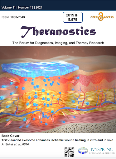
TGF-β loaded exosome enhances ischemic wound healing in vitro and in vivo
With over seven million infections and $25 billion treatment cost, chronic ischemic wounds are one of the most serious complications in the United States. The controlled release of bioactive factor enriched exosome from fibrin gel was a promising strategy to promote wound healing.
Theranostics | April 30, 2021
TGF-β loaded exosome enhances ischemic wound healing in vitro and in vivo
With over seven million infections and $25 billion treatment cost, chronic ischemic wounds are one of the most serious complications in the United States. The controlled release of bioactive factor enriched exosome from finbrin gel was a promising strategy to promote wound healing.
Biotechnol Bioengineering | August 4, 2023
Dose-response analysis after administration of a human platelet-derived exosome product on neurite outgrowth in vitro
Modulating the nerve’s local microenvironment using exosomes is proposed to enhance nerve regeneration. This study aimed to determine the optimal dose of purified exosome product (PEP) required to exert maximal neurite extension. An in vitro dorsal root ganglion (DRG) neurite outgrowth assay was used to evaluate the effect of treatment with (i) 5% PEP, (ii) 10% PEP, (iii) 15% PEP, or (iv) 20% PEP on neurite extension (N = 9/group), compared to untreated controls. After 72 h, neurite extension was measured to quantify nerve regeneration. Live cell imaging was used to visualize neurite outgrowth during incubation. Treatment with 5% PEP resulted in the longest neurite extension and was superior to the untreated DRG (p = 0.003). Treatment with 10% PEP, 15% PEP, and 20% PEP was found to be comparable to controls (p = 0.12, p = 0.06, and p = 0.41, respectively) and each other. Live cell imaging suggested that PEP migrated towards the DRG neural regeneration site, compared to the persistent homogenous distribution of PEP in culture media alone. 5% PEP was found to be the optimal concentration for nerve regeneration based on this in vitro dose–response analysis.
Plastic and Reconstructive Surgery | March 14, 2023
Enhancing Functional Recovery after Segmental Nerve Defect using Nerve Allograft treated with Plasma-Derived Exosome
Nerve injuries can result in detrimental functional outcomes. Currently, autologous nerve graft offers the best outcome for segmental peripheral nerve injury. Allografts are alternatives, but do not have comparable results. This study evaluated whether plasma-derived exosome can improve nerve regeneration and functional recovery when combined with decellularized nerve allografts.
Regenerative Medicine, Vol. 18, NO. 1 | October 18, 2022
Purified exosome product enhances chondrocyte survival and regeneration by modulating inflammatory and promoting chondrogenesis.
This study was to detect the effects of purified exosome product (PEP) on C28/I2 cells and chondrocytes derived from osteoarthritis patients.
Discovery’s Edge | October 12, 2022
Early Research Toward a Cell-Free Solution for Stress Urinary Incontinence
The research team used regenerative purified exosome product, known as PEP, derived from platelets to deliver messages into the cells of preclinical models. Exosomes are extracellular vesicles that are like a delivery service moving cargo from one cell to another, with instructions for targeting exact tissues that need repair. The study suggests that the use of purified exosome product alleviates stress urinary incontinence from musculoskeletal breakdown in animals. The team did not detect any infection or off-target toxicity with application of PEP
Discovery’s Edge| October 12, 2022
Early Research Toward a Cell-Free Solution for Stress Urinary Incontinence
The research team used regenerative purified exosome product, known as PEP, derived from platelets to deliver messages into the cells of preclinical models. Exosomes are extracellular vesicles that are like a delivery service moving cargo from one cell to another, with instructions for targeting exact tissues that need repair. The study suggests that the use of purified exosome product alleviates stress urinary incontinence from musculoskeletal breakdown in animals. The team did not detect any infection or off-target toxicity with application of PEP
Regenerative Medicine, Vol. 17, NO. 11 | October 4, 2022
Platelet-derived Exosomes Induce Cell Proliferation and Wound Healing in Human Endormetrial Cells.
Aim: To investigate the regenerative effects of a platelet-derived purified exosome product (PEP) on human endometrial cells. Materials & methods: Endometrial adenocarcinoma cells (HEC-1A), endometrial stromal cells (T HESC) and menstrual blood-derived stem cells (MenSC) were assessed for exosome absorption and subsequent changes in cell proliferation and wound healing properties over 48 h. Results: Cell proliferation increased in PEP treated T HESC (p < 0.0001) and MenSC (p < 0.001) after 6 h and in HEC-1A (p < 0.01) after 12 h. PEP improved wound healing after 6 h in HEC-1A (p < 0.01) and MenSC (p < 0.0001) and in T HESC between 24 and 36 h (p < 0.05). Conclusion: PEP was absorbed by three different endometrial cell types. PEP treatment increased cell proliferation and wound healing capacity.
NJP Regen. Med | September 29, 2022
Exosome biopotentiated hydrogel restores damaged skeletal muscle in a porcine model of stress urinary incontinence.
Urinary incontinence afflicts up to 40% of adult women in the United States. Stress urinary incontinence (SUI) accounts for approximately one-third of these cases, precipitating ~200,000 surgical procedures annually. Continence is maintained through the interplay of sub-urethral support and urethral sphincter coaptation, particularly during activities that increase intra-abdominal pressure. Currently, surgical correction of SUI focuses on the re-establishment of sub-urethral support. However, mesh-based repairs are associated with foreign body reactions and poor localized tissue healing, which leads to mesh exposure, prompting the pursuit of technologies that restore external urethral sphincter function and limit surgical risk. The present work utilizes a human platelet-derived CD41a and CD9 expressing extracellular vesicle product (PEP) enriched for NF-κB and PD-L1 and derived to ensure the preservation of lipid bilayer for enhanced stability and compatibility with hydrogel-based sustained delivery approaches. In vitro, the application of PEP to skeletal muscle satellite cells in vitro drove proliferation and differentiation in an NF-κB-dependent fashion, with full inhibition of impact on exposure to resveratrol. PEP biopotentiation of collagen-1 and fibrin glue hydrogel achieved sustained exosome release at 37 °C, creating an ultrastructural “bead on a string” pattern on scanning electron microscopy. Initial testing in a rodent model of latissimus dorsi injury documented activation of skeletal muscle proliferation of healing.
Orthopaedic Journal of Sports Medicine| December 2021
Intrinsic Tendon Regeneration After Application of Purified Exosome Product: An In Vivo Study.
Female Pelvic Medicine & Reconstructive Surgery | October 2021
(FPMRS) Exosome-induced vaginal tissue regeneration in a porcine mesh exposure model.
The purpose of this study was to explore the utility of an injectable purified exosome product derived from human apheresis blood to (1) augment surgical closure of vaginal mesh exposures, and (2) serve as a stand-alone therapy for vaginal mesh exposure.
Biomaterials | September 2021
Effects of purified exosome product on rotator cuff tendon-bone healing in vitro and in vivo.
Exosomes have multiple therapeutic targets, but the effects on healing rotator cuff tear (RCT) remain unclear. As a circulating exosome, purified exosome product (PEP) has the potential to lead to biomechanical improvement in RCT. Here, we have established a simple and efficient approach that identifies the function and underlying mechanisms of PEP on cell-cell interaction using a co-culture model in vitro.
Plastic and Reconstructive Surgery | August 2021
Administration of purified exosome product in a rat sciatic nerve reverse autograft model.
The nerve autograft remains the gold standard when reconstructing peripheral nerve defects. However, although autograft repair can result in useful functional recovery, poor outcomes are common, and better treatments are needed. The purpose of this study was to evaluate the effect of purified exosome product on functional motor recovery and nerve-related gene expression in a rat sciatic nerve reverse autograft model.
Female Pelvic Medicine & Reconstructive Surgery | March 1, 2021
(FPMRS) Impact of repeat dosing and mesh exposure chronicity on exosome-induced vaginal tissue regeneration in a porcine mesh exposure model.
The aim of the study was to compare vaginal wound healing after exosome injection in a porcine mesh exposure model with (1) single versus multiple dose regimens and (2) acute versus subacute exposure.
Regen Med | February 24, 2021
(Review) Regenerative medicine clinical readiness.
Regenerative medicine, poised to transform 21st century healthcare, has aspired to enrich care options by bringing cures to patients in need. Science-driven responsible and regulated translation of innovative technology has enabled the launch of previously unimaginable care pathways adopted prudently for select serious diseases and disabilities. The collective resolve to advance the design, manufacture and validity of affordable regenerative solutions aims to democratize such health benefits for all. The objective of this Review is to outline the framework and prerequisites that underpin clinical readiness of regenerative care. Integrated research and development, specialized workforce education and accessible evidence-based practice implementation are at the core of realizing an equitable regenerative medicine vision.
Journal of Orthopaedic Research | September 16, 2020
A novel engineered purified exosome product patch for tendon healing: An explant in an ex vivo model.
Reducing tendon failure after repair remains a challenge due to its poor intrinsic healing ability. The purpose of this study is to investigate the effect of a novel tissue-engineered purified exosome product (PEP) patch on tendon healing in a canine ex vivo model. Lacerated flexor digitorum profundus (FDP) tendons from three canines’ paws underwent simulated repair with Tisseel patch alone or biopotentiated with PEP. For the ex vivo model, FDP tendons were randomly divided into three groups: FDP tendon repair alone group (Control), Tisseel patch alone group, and the Tisseel plus PEP (TEPEP) patch group. Following 4 weeks of tissue culture, the failure load, stiffness, histology, and gene expression of the healing tendon were evaluated. Transmission electron microscopy revealed that in exosomes of PEP the diameters ranged from 93.70 to 124.65 nm, and the patch release test showed this TEPEP patch could stably release the extracellular vesicle over 2 weeks. The failure strength of the tendon in the TEPEP patch group was significantly higher than that of the Control group and Tisseel alone group. The results of histology showed that the TEPEP patch group had the smallest healing gap and the largest number of fibroblasts on the surface of the injured tendon. Quantitative reverse transcription polymerase chain reaction showed that TEPEP patch increased the expression of collagen type III, matrix metallopeptidase 2 (MMP2), MMP3, MMP14, and reduced the expression of transforming growth factor β1, interleukin 6. This study shows that the TEPEP patch could promote tendon repair by reducing gap formation and inflammatory response, increasing the activity of endogenous cells, and formation of type III collagen.
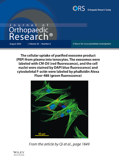
Journal of Orthopaedic Research | January 12, 2020
Characterization of a purified exosome product and its effects on canine flexor tenocyte biology.
Flexor tendon injuries and tendinopathy are very common but remain challenging in clinical treatment. Exosomes-based cell-free therapy appears to be a promising strategy for tendon healing, while limited studies have evaluated its impacts on tenocyte biology. The objective of this study was to characterize a novel purified exosome product (PEP) derived from plasma, as well as to explore its cellular effects on canine tenocyte biology. The transmission electron microscope revealed that exosomes of PEP present cup-shaped structures with the diameters ranged from 80 to 141 nm, and the NanoSight report presented that their size mainly concentrated around 100 nm. The enzyme-linked immunosorbent assay kits analysis showed that PEP was positive for CD63 and AChE expression, and the cellular uptake of exosomes internalized into tenocyte cytoplasm was observed. The cell growth assays displayed that tenocyte proliferation ability was enhanced by PEP solution in a dose-dependent manner. Tenogenic phenotype was preserved as is evident by that tendon-related genes expression (SCX, COL1A, COL3A1, TNMD, DCN, and MKX) were expressed insistently in a high level, while tenocytes were treated with 5% PEP solution. Furthermore, migration capability was maintained and total collagen deposition was increased. More interesting, dexamethasone-induced cellular apoptosis was attenuated during the incubation of tenocytes with a 5% PEP solution. These findings will provide the basic understandings about the PEP, and support the potential use of this biological strategy for tendon healing.
European Heart Journal | April 1, 2019
(Review) Regeneration for All: An Odyssey in Biotherapy: Authors from the Mayo Clinic discuss the evolution of the regenerative paradigm and look to the future.
Two decades ago, if you were to ask a scientist what it would mean to regenerate the heart, you would regularly hear about the power of developmental biology in decoding the intricacy of organogenesis and the plasticity of stem cells recognized as nature’s ultimate ‘building blocks’. The conversation would then delve into the merits and drawbacks of embryonic stem cell technology, presented as the quintessential regenerative phenotype.
With an ever-broader scholarly engagement and a growing public awareness, the academic intrigue of stem cell-based therapy was exponentially fuelled by the practicality of mining adult stem cell reservoirs out of bone marrow and adipose tissue. Universally, this multinational transdisciplinary endeavour captured the imagination of patients and physicians/scientists alike, while recognizing the ensuing medical, ethical and societal opportunities, and potential risks.
The prospect of pioneering a change in disease management propelled this maturing field from science fiction to the rigor of the scientific bench and onwards to randomized clinical trials. Here, the tantalizing concept of rebuilding the body to reverse underlying pathology, as opposed to a battle to palliate disease, emerged as a paradigm shift. In parallel, the notion of a curative intervention held the promise of altering the economics of chronic disease management, relieving the health care system of the cost burden associated with an ageing population vulnerable to degenerative disease.
